Chung Lab News

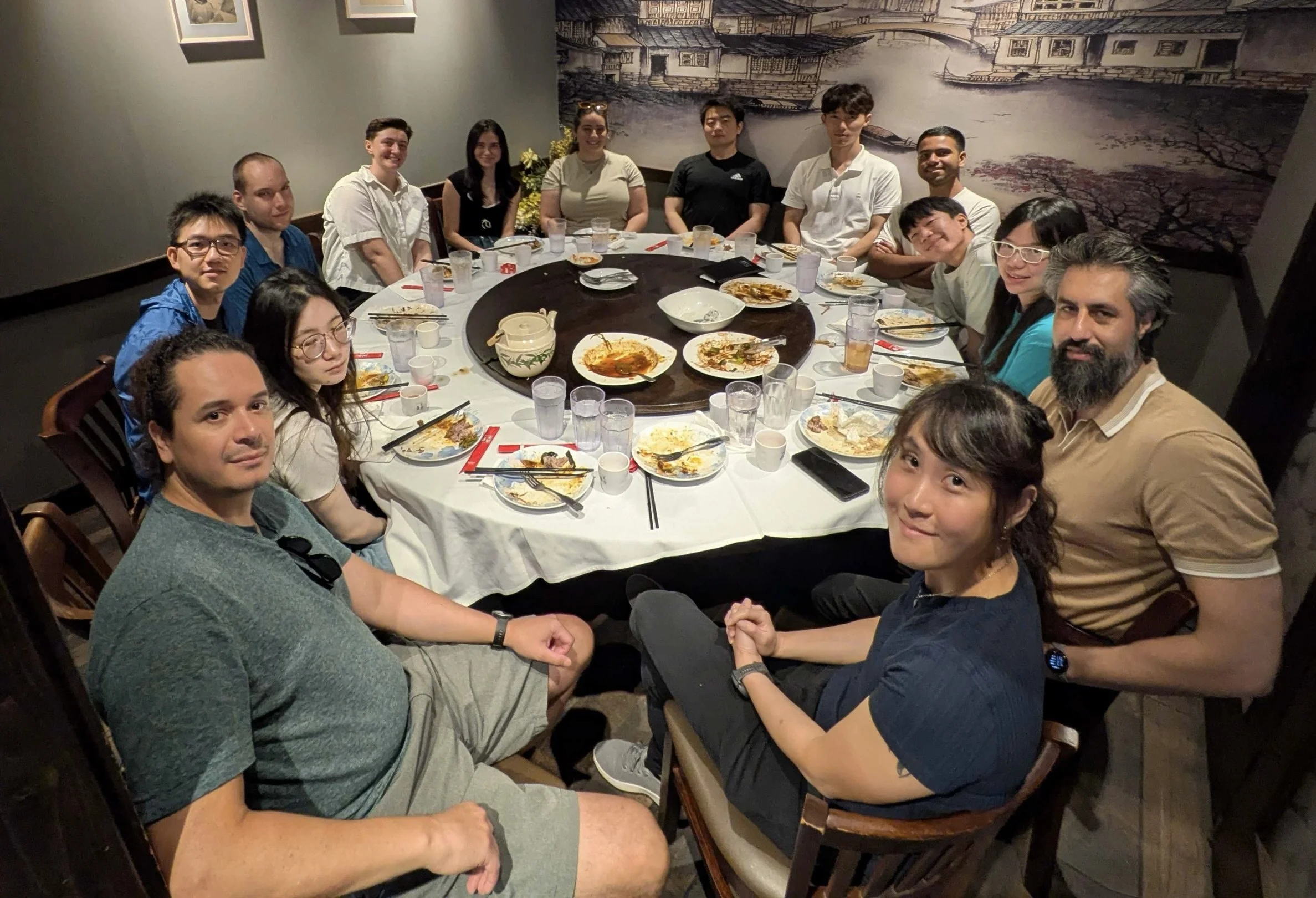
Welcome Lunch for our new Lab Members
Post Welcome Lunch at MuLan for our new Lab Members: Layla and Maddy!

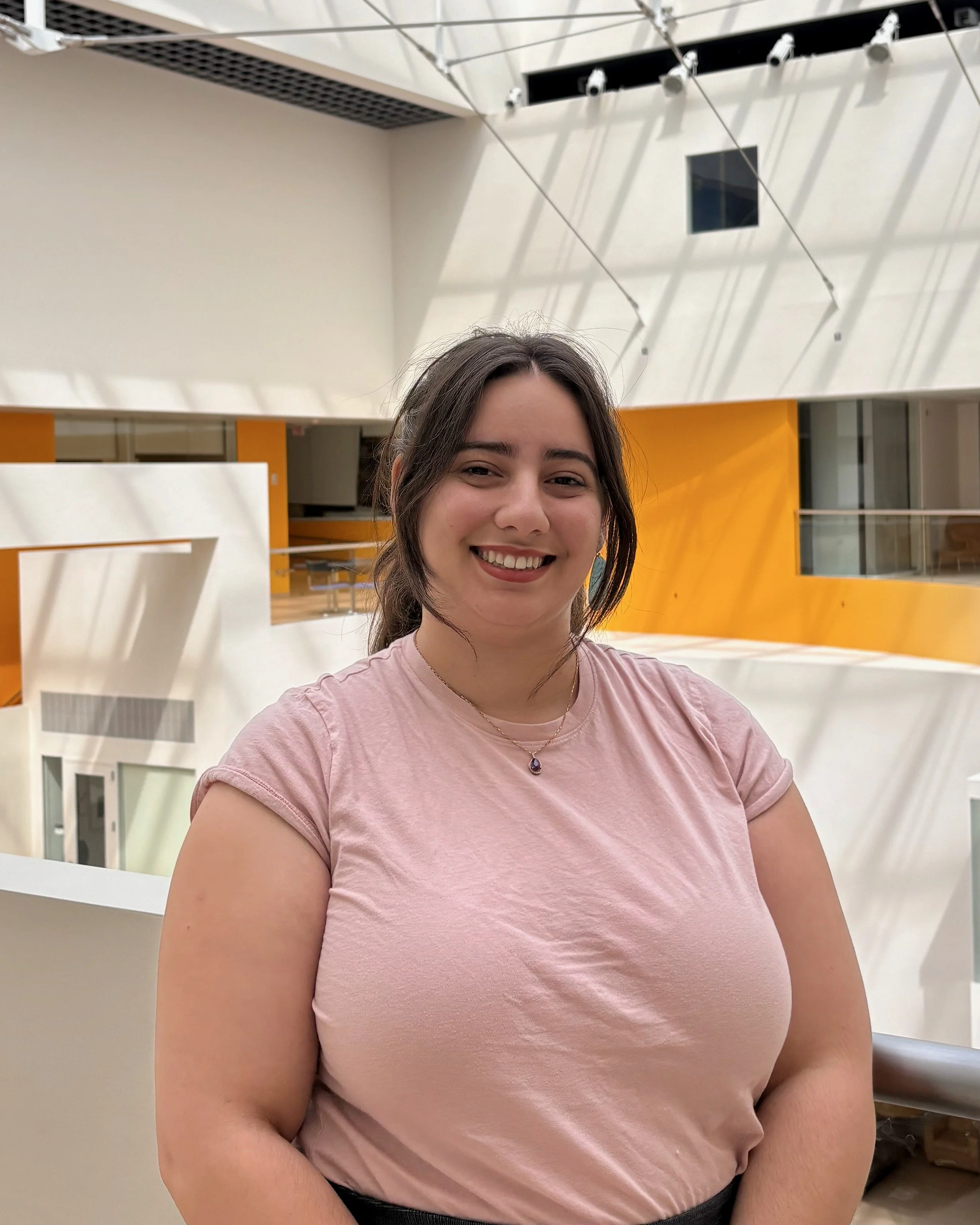
Welcome our New Lab Member: Madeline Hon!
Please join us in welcoming Madeline (Maddy) Hon to the Chung Lab!
Maddy will be pursuing her master’s thesis on the computational side of the UM1 project, with a focus on developing machine learning and computer vision models to analyze our datasets. We’re excited to have her on the team and look forward to the contributions she will make.
Mackenzie Awarded the Department of Chemistry Award for Outstanding Teaching
We are thrilled to announce that Mackenzie Riley has been honored with the Department of Chemistry Award for Outstanding Teaching. This award recognizes graduate students who have demonstrated exceptional dedication, creativity, and effectiveness in their teaching roles within the Chemistry Department.
Mackenzie received this recognition as a result of her excellent performance in teaching 5.111 Principles of Chemical Science in both Fall 2024 and Spring 2025. Her commitment to fostering an engaging and supportive learning environment has left a lasting impact on students and colleagues alike.
Please join us in celebrating Mackenzie for this well-deserved recognition!

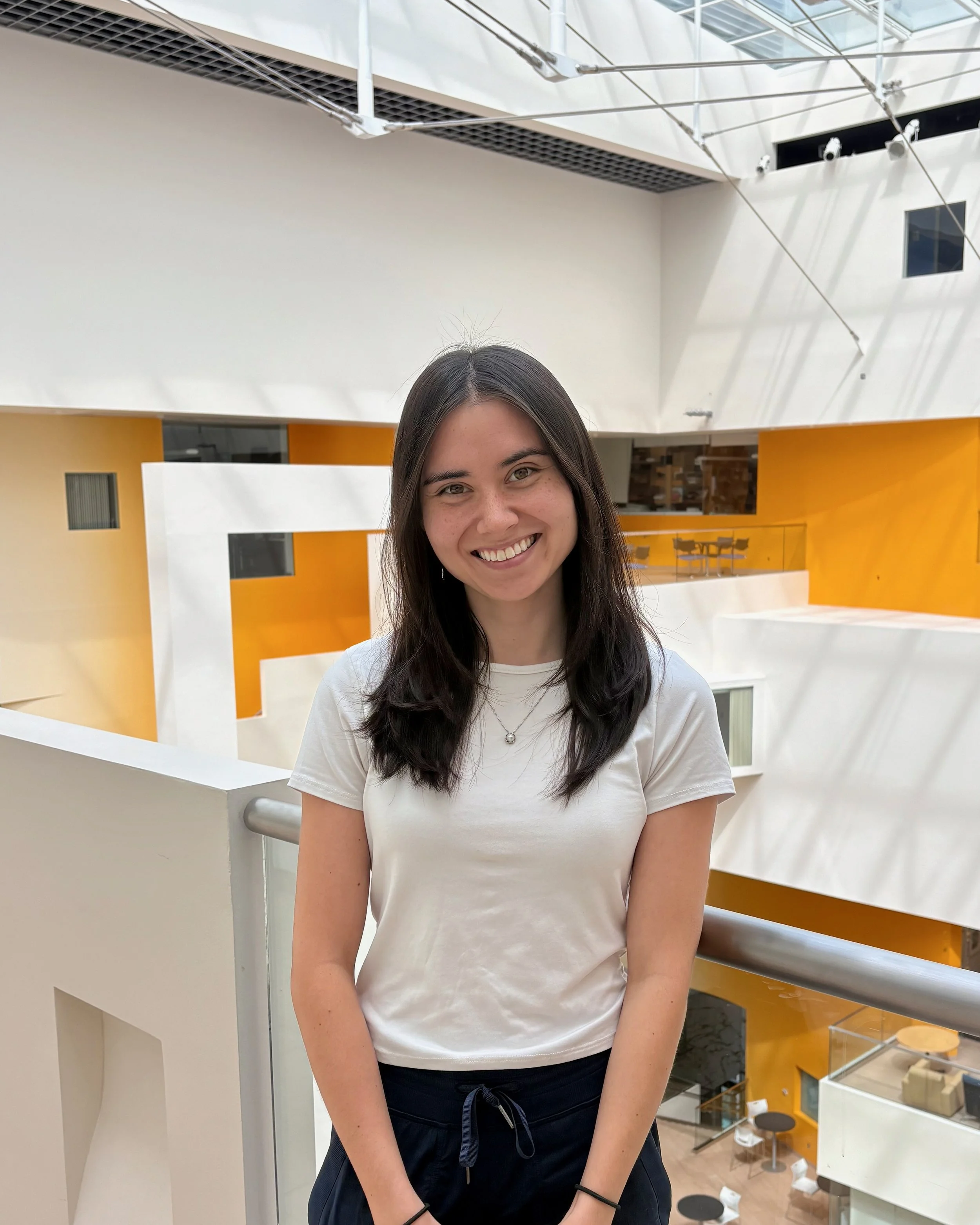
Welcome our New Administrative Assistant: Layla Gonzalez
Please join us in welcoming our new Administrative Assistant, Layla Gonzalez!
Layla joined the Chung Lab on August 18, 2025, and is already contributing her organizational skills and energy to the team. Originally from Miami, FL, she moved to Cambridge, MA in Fall 2023 and has been enjoying all that New England has to offer. Outside of work, Layla enjoys playing board games, reading, and exploring everything New England has to offer.
We are excited to have Layla on board and look forward to working with her!
Gokulnath Ganesan Selected for the MathWorks Fellowship
We are proud to announce that our lab member, Gokulnath Ganesan, has been selected as a MathWorks Fellow. This prestigious fellowship recognizes outstanding graduate students at MIT whose research leverages advanced computational tools such as MATLAB and Simulink.
The MathWorks Fellowship provides generous support to fellows as they pursue bold and innovative research directions. Awardees also gain mentorship and networking opportunities through MathWorks, strengthening the bridge between academic research and industry applications.
Gokul is a 2nd year PhD student in the Chung lab, developing minimally invasive devices for cell therapy delivery. The MathWorks Fellowship will support his efforts to create a MATLAB-integrated development pipeline that assists the design, simulation, and fabrication of therapeutic devices. This computational pipeline will greatly accelerate design iteration and support spatially tailored device architecture, which have been key bottlenecks in the early-stage development of cell therapy devices.
Please join us in congratulating Gokul for this remarkable achievement!
Keyu Chen Selected for the HEALS Fellowship
We are delighted to share that Keyu Chen has been awarded the HEALS (Health, Environment, and Life Sciences) Fellowship. The HEALS program is an interdisciplinary graduate fellowship at MIT that brings together students from across the Institute to address pressing challenges at the intersection of human health and the environment.
The fellowship supports research training that combines cutting-edge life sciences, data science, and environmental health approaches, preparing the next generation of scientists to tackle complex issues that impact both individuals and communities.
Keyu’s selection as a HEALS Fellow recognizes not only his academic excellence but also the broader impact of his research. Driven by a passion for interdisciplinary approaches to improve human health, he is using high-resolution printing integrated with stem cell cultures to design implantable devices for organ replacement and regenerative therapy.
Please join us in congratulating Keyu on this exciting achievement!

Chung lab postdoc honored with 2025 Koch Institute Image Award
One of the postdoctoral researchers from the Chung Lab, Dr. Lorenzo Ochoa, has been named a recipient of the 2025 Koch Institute Image Award; the award recognizes exceptional scientific images that are visually striking while conveying significant insights into biomedical research. The winning image showcases cutting-edge advancements in high-resolution brain mapping, a core focus of the lab’s research. This achievement highlights the lab’s commitment to developing innovative imaging techniques that deepen our understanding of brain structure and function. The award celebrates both the aesthetic and scientific impact of their work, reinforcing the importance of visualizing complex biological systems to drive discovery.

Chung Lab Featured in MIT News: Breakthrough Technologies for Protein Labeling of Intact Tissues
We are excited to share that our lab’s latest innovations have been featured in MIT News! The article showcases the new CuRVE and eFLASH technologies, which enable ultrafast and uniform protein labeling across tens of millions of densely packed individual cells in fully intact 3D tissues with unprecedented precision and scalability. This breakthrough addresses critical challenges in studying complex biological systems, such as brain tissue and organoids, where dense cellular environments often hinder detailed protein analysis. By integrating advanced molecular engineering, high-resolution imaging, and computational analysis, our approach allows for more accurate characterization of protein distributions in situ. This innovation significantly improves the speed and accuracy of protein visualization, unlocking new possibilities for biological and neuroscience research.


Dr. Juhyuk Park is heading off to a tenure faculty position at Seoul National University!
We are thrilled to announce that Dr. Juhyuk Park is heading off to a tenure faculty position at Seoul National University’s Materials Sciences and Engineering department!

Chung Lab member presents their work at the Tissue Clearing Workshop 2024
Our senior postdoc fellow, Dr Juhyuk Park, presented the Chung lab tissue processing pipeline on behalf of Prof Chung at the Tissue Clearing Workshop 2024, hosted by Harvard BioLabs, Cambridge, MA. Talk title: "Tissue clearing and processing technology for human brain mapping"

Welcome Lunch for Chung Lab's newest member, Dominik Kylies.
Please welcome our new member, Dominik Kylies!


